
[ad_1]
Abstract: Scientists have created a groundbreaking spatial cell atlas of the human limb, capturing the intricate technique of human limb growth. This work, a part of the Human Cell Atlas initiative, marks a major development in understanding the speedy and complicated formation of human limbs.
The research gives an in depth map of the mobile panorama throughout early limb formation, providing insights into congenital limb syndromes. This analysis not solely deepens our understanding of human anatomy but in addition opens avenues for diagnosing and treating varied limb-related problems.
Key Information:
- The atlas showcases how human fingers and toes type from a bigger foundational bud, with intervening cells receding to disclose the digits.
- This research is part of the worldwide Human Cell Atlas initiative, aiming to map each cell sort within the human physique to revolutionize our understanding of well being and illness.
- The atlas uncovers new hyperlinks between developmental cells and congenital limb syndromes, similar to brachydactyly (brief fingers) and polysyndactyly (further digits).
Supply: Wellcome Sanger Institute
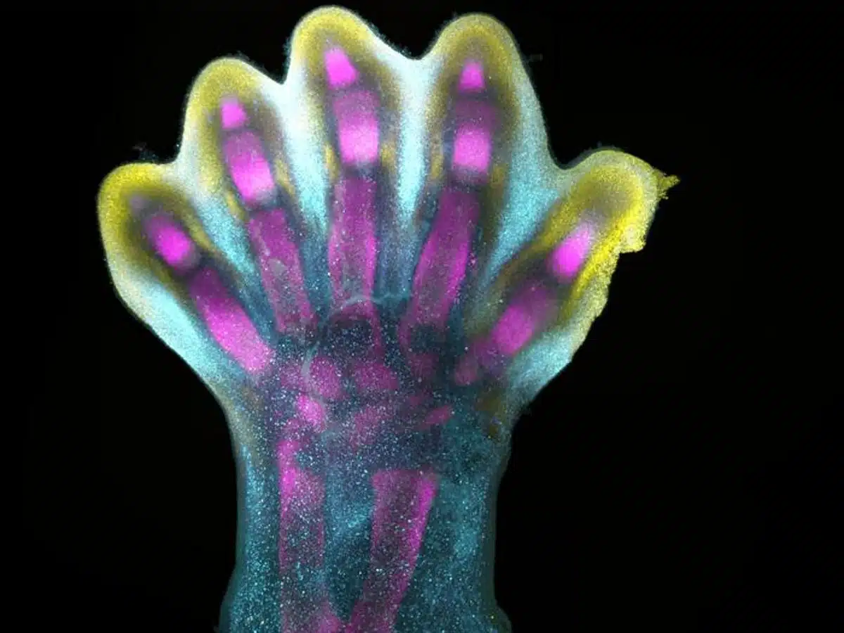
Human fingers and toes don’t develop outward; as a substitute, they type from inside a bigger foundational bud, as intervening cells recede to disclose the digits beneath. That is amongst many processes captured for the primary time as scientists unveil a spatial cell atlas of all the growing human limb, resolved in area and time.
Researchers on the Wellcome Sanger Institute, Solar Yat-sen College, EMBL’s European Bioinformatics Institute and collaborators utilized cutting-edge single-cell and spatial applied sciences to create an atlas characterising the mobile panorama of the early human limb, pinpointing the precise location of cells.
This research is a part of the worldwide Human Cell Atlas initiative to map each cell sort within the human physique, to rework understanding of well being and illness.
The atlas, revealed at present (6 December) in Nature, gives an overtly out there useful resource that captures the intricate processes governing the limbs’ speedy growth through the early phases of limb formation.
The atlas additionally uncovers new hyperlinks between developmental cells and a few congenital limb syndromes, similar to brief fingers and additional digits.
Limbs are recognized to initially emerge as undifferentiated cell pouches on the edges of the physique, and not using a particular form or operate. Nonetheless after 8 weeks of growth, they’re effectively differentiated, anatomically complicated and instantly recognisable as limbs, full with fingers and toes.
This requires a really speedy and exact orchestration of cells. Any small disturbances to this course of can have a downstream impact, which is why variations within the limbs are among the many most incessantly reported syndromes at beginning, affecting roughly one in 500 births globally.
Whereas limb growth has been extensively studied in mouse and chick fashions, the extent to which they mirror the human state of affairs remained unclear. Nonetheless, advances in know-how now allow researchers to discover the early phases of human limb formation.
On this new research, scientists from the Wellcome Sanger Institute, Solar Yat-sen College, and their collaborators analysed tissues between 5 and 9 weeks of growth. This allowed them to hint particular gene expression packages, activated at sure instances and in particular areas, which form the forming limbs.
Particular staining of the tissue revealed clearly how cell populations differentially organize themselves into patterns of the forming digits.
As a part of the research, researchers demonstrated that sure gene patterns have implications for a way the palms and ft type, figuring out sure genes, which when disrupted, are related to particular limb syndromes like brachydactyly – brief fingers – and polysyndactyly – further fingers or toes.
The staff had been additionally in a position to verify that many points of limb growth are shared between people and mice.
General, these findings not solely present an in-depth characterisation of limb growth in people but in addition essential insights that would impression the analysis and therapy of congenital limb syndromes.
Professor Hongbo Zhang, senior creator of the research from Solar Yat-sen College, Guangzhou, stated: “Many years of finding out mannequin organisms established the premise for our understanding of vertebrate limb growth. Nonetheless, characterising this in people has been elusive till now, and we couldn’t assume the relevance of mouse fashions for human growth.
“What we reveal is a extremely complicated and exactly regulated course of. It’s like watching a sculptor at work, chiselling away at a block of marble to disclose a masterpiece. On this case, nature is the sculptor, and the result’s the unimaginable complexity of our fingers and toes.”
Dr Sarah Teichmann, senior creator of the research from the Wellcome Sanger Institute, and co-founder of the Human Cell Atlas, stated: “For the primary time, now we have been in a position to seize the outstanding technique of limb growth right down to single cell decision in area and time.
“Our work within the Human Cell Atlas is deepening our understanding of how anatomically complicated constructions type, serving to us uncover the genetic and mobile processes behind wholesome human growth, with many implications for analysis and healthcare.
“As an example, we found novel roles of key genes MSC and PITX1 which will regulate muscle stem cells. This might supply potential for treating muscle-related problems or accidents.”
About this cell mapping and developmental neuroscience analysis information
Writer: Jelena Pupavac
Supply: Wellcome Sanger Institute
Contact: Jelena Pupavac – Wellcome Sanger Institute
Picture: The picture is credited to the researchers/Nature
Authentic Analysis: Open entry.
“A human embryonic limb cell atlas resolved in area and time” by Sarah Teichmann et al. Nature
Summary
A human embryonic limb cell atlas resolved in area and time
Human limbs emerge through the fourth post-conception week as mesenchymal buds, which turn into absolutely fashioned limbs over the following months. This course of is orchestrated by quite a few temporally and spatially restricted gene expression programmes, making congenital alterations in phenotype frequent.
Many years of labor with mannequin organisms have outlined the basic mechanisms underlying vertebrate limb growth, however an in-depth characterization of this course of in people has but to be carried out.
Right here we element human embryonic limb growth throughout area and time utilizing single-cell and spatial transcriptomics. We display in depth diversification of cells from just a few multipotent progenitors to myriad differentiated cell states, together with a number of novel cell populations.
We uncover two waves of human muscle growth, every characterised by totally different cell states regulated by separate gene expression programmes, and determine musculin (MSC) as a key transcriptional repressor sustaining muscle stem cell id.
Via meeting of a number of anatomically steady spatial transcriptomic samples utilizing VisiumStitcher, we map cells throughout a sagittal part of an entire fetal hindlimb. We reveal a transparent anatomical segregation between genes linked to brachydactyly and polysyndactyly, and uncover transcriptionally and spatially distinct populations of the mesenchyme within the autopod.
Lastly, we carry out single-cell RNA sequencing on mouse embryonic limbs to facilitate cross-species developmental comparability, discovering substantial homology between the 2 species.
[ad_2]